Abstract
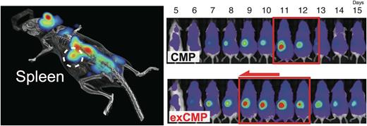
Hematopoietic stem cells (HSC) represent the apex of the hematopoietic hierarchy and their engraftment post-transplantation is essential for long-term donor hematopoiesis. After transplantation, injected HSCs rapidly expand to reconstitute the entire blood system. This early post-transplant "hematopoietic inflation" is a crucial phase in sustaining life. However, the time course of hematopoietic inflation and the spatial transition within the whole body have remained a mystery. The discovery of colonies forming in the spleen early after HSC transplantation in the 1960s has been a strong advocate of the existence of HSCs, but biological role of transient nature of spleen colonies has not been understood. In this study, we elucidated the spatiotemporal dynamics of hematopoietic inflation in the same individual in a murine HSC transplantation model. Current methods for tracking transplanted HSCs in vivo are limited due to their low sensitivity and their restriction to observe only previously known niches. To address these issues, we used the recently published, highly sensitive AkaLuc bioluminescence imaging system to visualize hematopoietic inflation in vivo. We combined Akaluc 3D images with CT scans to spatially capture early hematopoiesis after transplantation on a whole-body scale, and continuously monitored bioluminescence signals to clarify the dynamics of cellular distributions over time. Surprisingly, our data show that the spleen acts as a major site of hematopoiesis and shows unique hematopoietic patterns with dynamic changes. Splenic hematopoiesis was distinct from that occurring in the bone marrow and each progenitor cell has a different hematopoietic inflation pattern. We found that activated common myeloid progenitors (aCMPs) use the spleen as a site for hematopoietic inflation, producing much more cells early after transplantation than normal CMPs. Based on these findings, we developed expanded CMPs (exCMPs) that leverage the spleen environment to improve early hematopoietic recovery after transplantation. exCMPs, which can be produced in large quantities in polyvinyl-alcohol (PVA)-based cultures (Wilkinson et al., Nature 2019), had lower CXCR4 expression, different integrin expression patterns, better cytokine responsiveness and mitogenic potential, as well as higher expression of platelet-related genes compared to fresh CMP. Therefore, exCMPs are more likely to migrate to the spleen and proliferate more rapidly than fresh CMPs. Exploiting these properties, we could show that weekly administration of exCMP allowed for the long-term survival of lethally irradiated mice in a spleen-dependent manner. In the allogeneic transplantation setting, universal exCMPs could sustain life for a long time by a serial transplantation. Human exCMPs could be easily expanded as well, and the clinical use of human exCMPs is highly anticipated as a supportive therapy after HSCT. Our studies of post-transplant hematopoiesis using a next-generation luminescence imaging system showed that the spleen functioned as a major hematopoietic tissue and that aCMPs and the spleen cooperated to contribute to hematopoietic inflation post-SCT, which led to the concept of utilizing the spleen as an alternative hematopoietic site. These findings refine our understanding of the dynamics of hematopoietic reconstitution, reveal the unique collaboration of the spleen and transplanted cells and establish the therapeutic potential of exCMPs for early hematopoietic recovery.
Disclosures
No relevant conflicts of interest to declare.
Author notes
Asterisk with author names denotes non-ASH members.


This feature is available to Subscribers Only
Sign In or Create an Account Close Modal